Contact us!     1-877-355-8012  /  info@vielight.com

Vielight Logo
0

Your cart is currently empty.

Vielight Shop
0

Your cart is currently empty.

Vielight Logo
0

Your cart is currently empty.

Brain Imaging Studies with Vielight Neuro Reveal the Network Effects of Brain Photobiomodulation

For years, brain photobiomodulation (PBM) was often described in local terms: more blood flow here, less inflammation there, happier mitochondria everywhere.

But over the last few years, something deeper has been coming into focus.

Independent MRI and fMRI studies from the University of Sydney, the University of California San Francisco (UCSF), and the University of Utah are showing that PBM with Vielight technology doesn’t just act locally. It appears to reshape how large-scale brain networks behave – especially the Default Mode Network (DMN) and its partners in attention and cognitive control.

This post walks through three key imaging studies:

  1. Healthy adults – neuromodulation and DMN deactivation during a task (University of Sydney)
  2. Dementia/Alzheimer’s – perfusion and DMN connectivity at rest (University of California San Francisco & VA San Francisco)
  3. Traumatic brain injury – cerebellar network efficiency in ex-football players (University of Utah)
  4. Mild Cognitive Impairment (MRI) – Pending Publication (Preprint – University of Toronto)
  5. Traumatic brain injury (DTI) – Pending Publication (University of Utah)

Together, they outline a consistent story: Vielight PBM strengthens the efficiency of core neural networks.

⚠️ These are research findings, not medical advice.

A quick refresher: What is the Default Mode Network?

The Default Mode Network is a set of regions (including the posterior cingulate cortex, medial prefrontal cortex, and lateral parietal areas) that typically:

  • Are more active when the mind is resting, daydreaming, or internally focused, and

  • Should deactivate when we shift into demanding tasks.

In many neurological conditions — including Alzheimer’s disease and TBI — this network shows abnormal connectivity and dysregulated activity, such as reduced within-network coherence or failure to deactivate appropriately during tasks.

That’s why DMN behavior is such a central theme in the PBM imaging story.

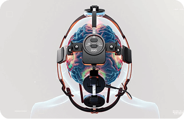
The Vielight Neuro 4’s geometry is engineered to intentionally overlap these broadened fields over Default Mode Network (DMN) nodes with the highest measured irradiance in commercially available brain photobiomodulation devices, while still bathing the wider cortex. This is why five VieLED modules can produce an effect that is effectively full‑transcranial, with a focus on the DMN.

Imaging Study 1 (MRI): University of Sydney: DMN deactivation in healthy young adults

Study: (Link)
El Khoury, H., Mitrofanis, J., & Henderson, L. A. (2019).
“Exploring the Effects of Near Infrared Light on Resting and Evoked Brain Activity in Humans Using Magnetic Resonance Imaging” (Neuroscience).

Device & protocol

  • Technology: Vielight Neuro

  • Population: 24 Normal, young adults.

  • Design: Participants underwent active vs sham PBM sessions, then performed fMRI at rest and during a finger-tapping task.

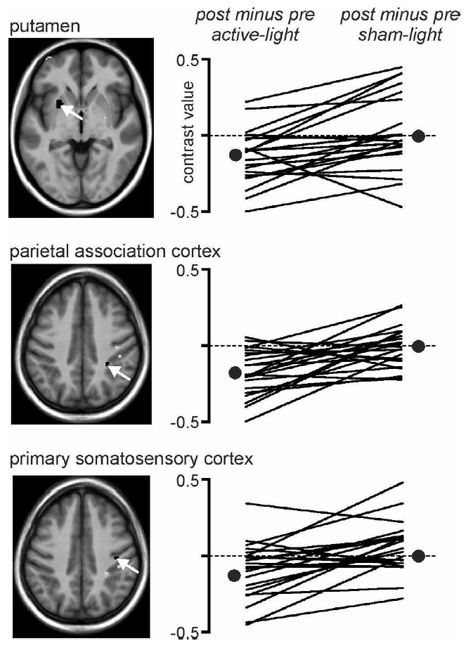
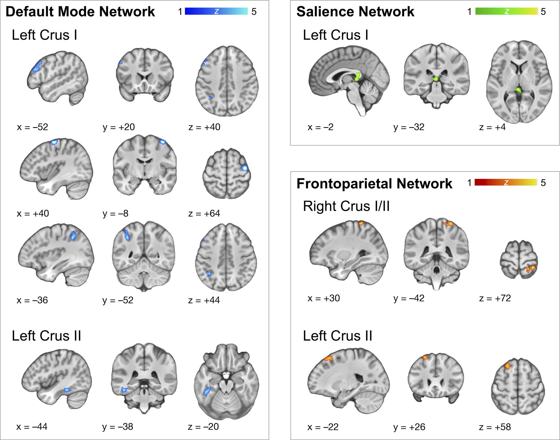
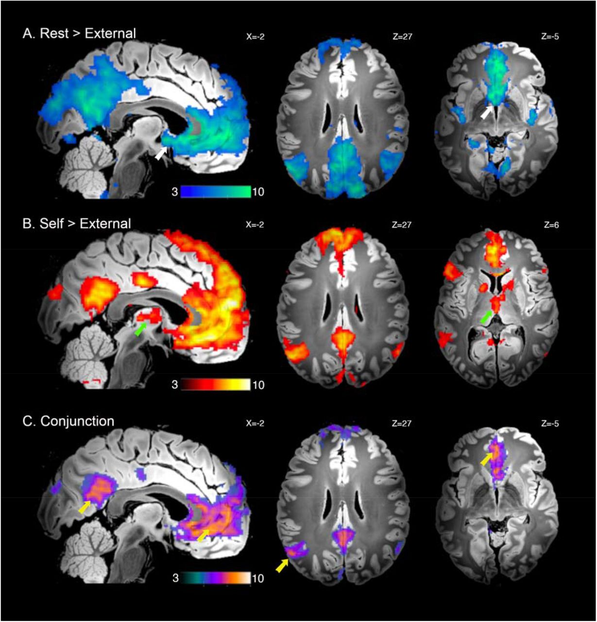
Figure 1. Active vs Sham - Vielight Neuro Effects of active- and sham-light on finger-tapping evoked activity. (A) Significant differences in finger-tapping evoked activation before and after active-light

At the University of Sydney, researchers asked an important question: if healthy young adults receive a single session with a Vielight 810 nm transcranial device, what changes on fMRI at rest and during a simple finger-tapping task?

Their answer: resting global activity didn’t budge, but during the task there were reductions in activity across regions you want to quiet when focusing (primary somatosensory cortex, parietal association cortex, putamen). Functional connectivity from parietal association cortex shifted as well. The team interprets these shifts as attention/novelty related and notes they “served to deactivate the so-called default mode network (DMN).”

Why that matters: in a healthy brain, the DMN should dial down when you focus. This study reads as PBM acting like context-sensitive gain control—not revving a resting brain, but helping it suppress background chatter the moment work begins.

The effect of active- and sham-light on individual subject brain activation.

Key imaging findings

  • During the finger-tapping task (a “task-positive” condition), PBM reduced task-evoked activity in:

    • Putamen

    • Primary somatosensory cortex

    • Parietal association cortex

  • Resting-state connectivity was largely unchanged, except for a specific change in the functional connectivity of the parietal association cortex.

The authors interpret these changes as being related to attention and novelty, and notably suggest that the pattern of activity “served to deactivate the so-called default mode network.”

What this means in plain language

  • The brain at rest doesn’t get randomly “amped up” by PBM.

  • Instead, when the brain needs to perform a task, PBM appears to help quiet down DMN-related regions that shouldn’t dominate during focused behavior.

In other words, in healthy brains PBM looks less like a “volume knob” and more like a precision gain control: enabling better suppression of default-mode processes when it’s time to work.

Imaging Study 2 (fMRI): Perfusion and DMN connectivity in dementia

Study (Alzheimer’s/dementia imaging): (Link)
University of California San Francisco, Chao, L. L. (2019).
“Effects of Home Photobiomodulation Treatments on Cognitive and Behavioral Function, Cerebral Perfusion, and Resting-State Functional Connectivity in Patients with Dementia: A Pilot Trial” – UCSF and San Francisco VA Medical Center.

This is the UCSF fMRI study highlighted in Vielight’s article “Alzheimer’s Disease and Brain Photobiomodulation | Clinical Results with Vielight Neuro.”

Figure 1 fMRI with Vielight Neuro

Increased cerebral perfusion with the Vielight Neuro Gamma

In a randomized clinical study at the University of California San Francisco and the San Francisco VA, eight older adults (average age ~80) were split into two equal groups. Four continued with usual care, while the other four added home photobiomodulation using the Vielight Neuro Gamma. The PBM group used the device three times per week for 12 weeks, with each session done at home in about 20 minutes.

To see whether the brain and behavior changed, the team collected two kinds of MRI and standard clinical measures before starting and after 12 weeks. First, they ran arterial spin labeling (ASL) perfusion MRI, a noninvasive way to estimate cerebral blood flow—think of it as a map of how well different brain regions are being supplied. Second, they ran resting-state fMRI, which tracks the natural, moment-to-moment synchrony between brain regions to assess the strength of large-scale networks like the Default Mode Network (DMN). Alongside the scans, participants completed cognitive tests and behavioral symptom scales, so the imaging could be interpreted next to real-world outcomes.

Figure 2. Arterial spin-labeled perfusion


Arterial spin-labeled perfusion values, normalized to the precentral ROI, from the superior frontal (A), superior parietal

Figure 3. ADAS-Cog and NPI Values

Mean (±SEM) ADAS-Cog (A) and NPI-FS (B) in three Usual Care patients before vs. after home PBM (blue). Lower scores = better function.

Clinical outcomes

After 12 weeks, the PBM group (vs usual care) showed:

  • Improved cognition

    • Significant gains on ADAS-cog (p ≈ 0.007)

  • Reduced behavioral symptoms

    • Significant improvements on Neuropsychiatric Inventory (NPI) scores

Importantly, no adverse effects were reported.

UCSF-Vielight Alzheimer's Imaging outcomes

The imaging adds the mechanistic “why”:

  1. Increased cerebral perfusion

    • PBM was associated with higher cerebral blood flow compared to usual care.

    • The increases were particularly notable in parietal regions, which matches the fact that the Neuro Gamma places three transcranial LED clusters over the parietal lobe and one over the frontal lobe.

  2. Increased DMN connectivity

    • Resting-state fMRI showed increased connectivity between the posterior cingulate cortex (PCC) and lateral parietal nodes of the Default Mode Network in the PBM group vs usual care.

    • That’s notable because disrupted DMN connectivity (especially involving the PCC) is a well-known feature of Alzheimer’s disease and at-risk populations.

How this complements the Sydney study

  • In healthy young adults, PBM helped deactivate DMN-related regions during a task.

  • In dementia, where DMN resting-state connectivity is typically impaired, PBM is associated with stronger intrinsic connectivity within the DMN and better perfusion, alongside improved cognitive and behavioral scores.

So in one case PBM supports “off when needed”; in the other, it supports “connected when needed.” The common theme is normalization, not simply “more activity is better.”

Imaging Study 3 (fMRI). Cerebellar network efficiency after repetitive head impacts

Study: (Link)
Wilde et al. (2025), University of Utah’s Traumatic Brain Injury and Concussion Center – summarized in Vielight’s “TBI Research Summary: Cerebellar Network Efficiency Improved by Vielight Intranasal-Transcranial Photobiomodulation (itPBM) (n=30)” and published in Photobiomodulation, Photomedicine, and Laser Surgery.

The University of Utah group studied adults with a history of repetitive head acceleration events – many with chronic post-impact symptoms. Participants used the Vielight Neuro, 20 min every other day for 8–10 weeks over the Default Mode Network (DMN). Resting-state fMRI focused on 11 cerebellar seed regions spanning DMN, Salience (SN), and Frontoparietal (FPN) networks.

Results: Within-network connectivity increased, while between-network connectivity decreased, a hallmark of higher network segregation/efficiency. Or in other terms, less noisy cross-talk, more coherent signaling inside the networks that matter for attention and control

Utah-Vielight TBI fMRI Imaging Results

Sample-specific seed-based connectivity maps for each region of interest are overlaid on an array of axial images in MNI template space, lateral–posterior–inferior (LPI) oriented, and represented in neurological view (left = left).

Imaging: Resting-state fMRI before and after the PBM course.

    • Analysis focused on 11 cerebellar seed regions linked to:

      • Default Mode Network (DMN)

      • Salience Network (SN)

      • Frontoparietal Network (FPN).

Key imaging findings

After the PBM intervention, the team found:

  1. Decreased between-network connectivity

    • Less “noisy crosstalk” between major networks — DMN, SN, and FPN.

  2. Increased within-network connectivity

    • Stronger internal coherence within these networks, especially Salience and Frontoparietal networks, which are crucial for attention, cognitive control, and sensory integration.

  3. Improved network segregation

    • The pattern of less between-network and more within-network connectivity suggests higher network efficiency – the networks appear more specialized and less diffusely entangled.

The authors and Vielight summary interpret this as evidence that PBM via the Neuro Gamma helps the brain regulate overactive, diffuse connections while strengthening the integrity of key, task-relevant networks.

In the context of RHAE/TBI — where resting-state connectivity can be pathologically altered — this provides a plausible neural mechanism to explain previously reported improvements in balance, motor function, and cognitive performance seen in related Utah studies using the same device.

Putting it all together: A network-level view of PBM

One mechanism, three stories

Taken together, these imaging results tell a consistent story even though the populations and outcomes differ:

  • Healthy brains: during tasks, PBM helps turn down DMN-like activity so task-relevant circuits can dominate.

  • Alzheimer’s/dementia: at rest, PBM is associated with stronger DMN connectivity and better perfusion where it’s often lacking, paralleling cognitive and behavioral gains in a small pilot.

  • Post-impact brains: PBM correlates with cleaner network boundaries—less random cross-talk, more internal coherence—which maps onto the kinds of attentional and executive symptoms people report after repetitive head impacts.

The throughline is regulation. PBM with Vielight technology doesn’t appear to simply excite tissue. It seems to help large-scale systems behave more appropriately for the context—idle when idling, engaged when engaging.

Where we go from here

These are promising but early findings: sample sizes are modest, and the field still needs larger randomized trials to confirm effects, tune protocols (wavelengths, pulse rates, session schedules), and identify who benefits most. That said, the imaging results already push the conversation beyond anecdotes. They show measurable changes in perfusion and functional connectivity that plausibly explain clinical gains reported across Alzheimer’s and brain-injury studies—and they align with a broader literature linking DMN behavior to real-world cognition. PubMed+2Liebert Publications+2

Imaging suggests PBM helps the brain organize itself. In healthy adults it supports DMN deactivation during tasks; in Alzheimer’s it supports DMN integration and perfusion at rest; and after repetitive head impacts it improves network efficiency by reducing cross-talk and tightening coherence.

Results Summary

  • Healthy brains didn’t show a blanket “activation,” but more effective DMN deactivation during a task—exactly what good attentional control requires.

  • Alzheimer’s/dementia brains, where DMN hubs and perfusion are often degraded, showed stronger DMN connectivity and increased CBF, with ADAS-Cog and NPI improvements in the PBM arm.

  • Post-impact brains showed cleaner network boundaries↑ within-network, ↓ between-network connectivity—consistent with more efficient large-scale communication.

That’s not a “more is better” story. It’s a normalization story: PBM with Vielight appears to help the brain organize itself—dialing down DMN when focus is needed, re-knitting fragile DMN links in disease, and reducing noisy cross-talk after injury.

This article was written by

Dr. Genane Loheswaran

Vielight | Research Manager and Neuroscientist

Genane manages Vielight’s research projects with various organizations for cognitive science, such as combining photobiomodulation with EEG.

MSc in Neuroscience, McMaster University
PhD in Pharmacology, University of Toronto
Meet the author
0